Noise-Field Perimetry (Noise-Field Test) Self-Check for Visual Field Impairment and Glaucoma as well as Early Detection of Glaucoma
Noise-field perimetry (noise-field test) is a simple test to create awareness of any visual field abnormalities a patient may have. Many patients with visual field defects due to various diseases have no subjective symptoms. In particular, patients with glaucoma may not experience subjective symptoms until the disease reaches an advanced stage. However, when watching noise-moving images, almost any patient can recognize abnormal areas, such as those with “no or little flicker,” “cloudy appearance,” or “blackish, gray, whitish appearance” that are consistent with visual field defects.
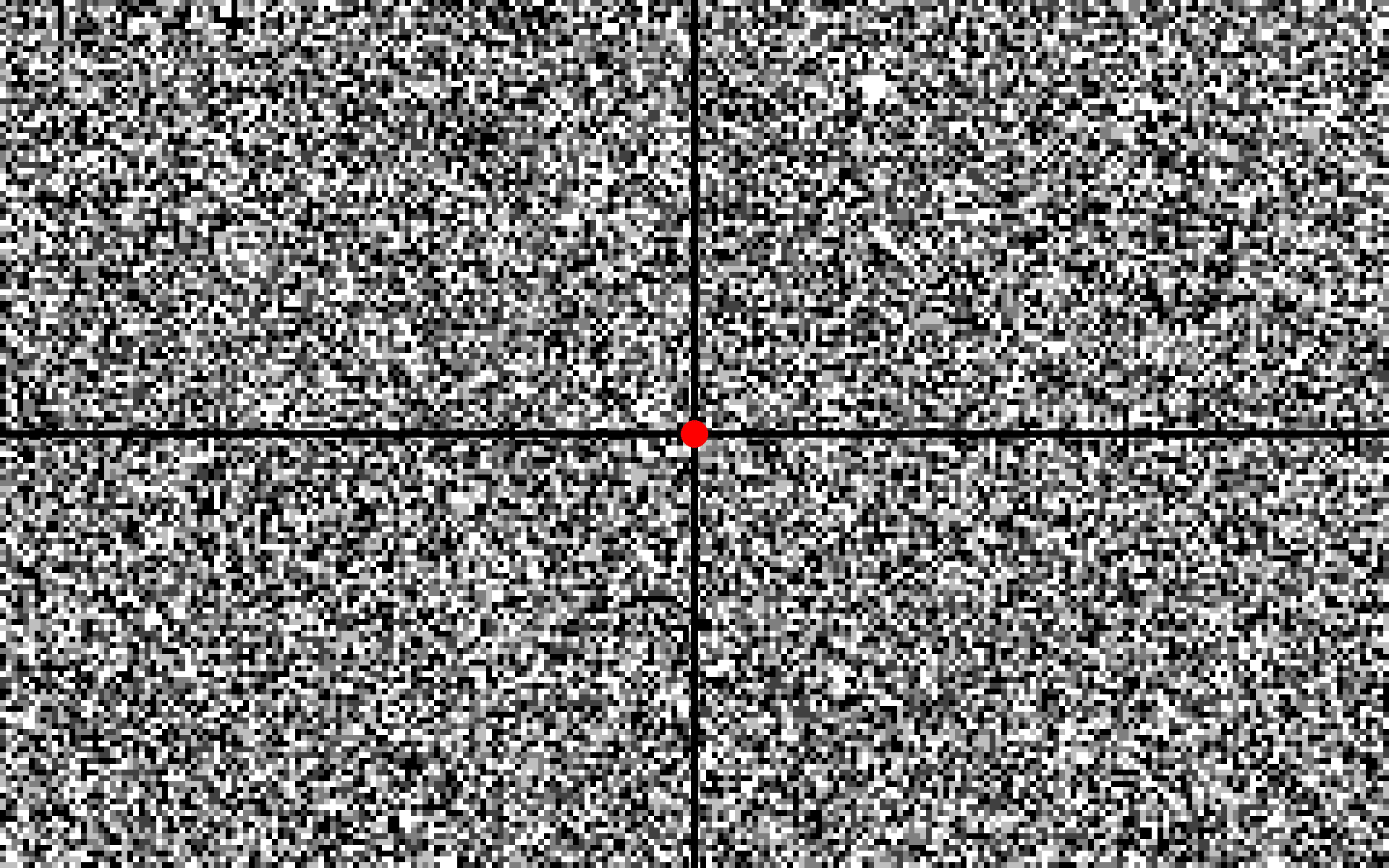
For this test, the patient simply watches a noise-moving image.
To a normal eye, nothing specific is visible; however, in the presence of a visual field defect, the patient will identify differences in the image within a few seconds.
The test may reveal abnormalities of the brain in addition to the abnormalities of the eyes.
Note:This is a still image.
A moving image is produced by the presentation of a sequence of still images.
Keyboard functions:
【←】【→】
:Adjust the size of the noise particles.
【↑】
:Show a white still image.
【↓】
:Show a black still image.
【Delete】
:Exit.
×
The primary purpose of this web test is to help patients understand their medical condition.
Even in patients with a disease, the web test may not detect any abnormality.
Whether the results of the web test show abnormalities or not, please be sure to consult an ophthalmologist if you have any concerns.
Do you consent to begin the test?
No
Having consented, please select your display screen type:
※Not available for smartphones.
Ophthalmologic Examination Guide, 3rd ed. Bunkodo, Tokyo: pp. 287-290, 2022, in Japanese.
A section of “noise-field campimetry” has been written by Dr. Arata Inoue, the director of Inoue Eye Clinic.
Test procedure
Preparation:
Ensure that the room is dark. In particular, no light should be focused on the display screen.
Recommended computer and display screen settings (not mandatory):
If possible, use a high-performance computer. On older computers, the test image will be less noise-moving; that is, it will flicker more slowly.
A larger display is preferable; 20 inches or larger is recommended.
Set the display screen brightness to its maximum.
Test start:
Ensure that the distance from your eyes to the display screen matches the display screen height.
The distance from your eyes to the display screen should match the display screen height.
Gaze at the red point in the image center (the fixation point) using one eye at a time. Cover the other eye with your hand.
Note: No distinction exists between the right and left eye for the noise screen. You may begin with either the right or the left eye.
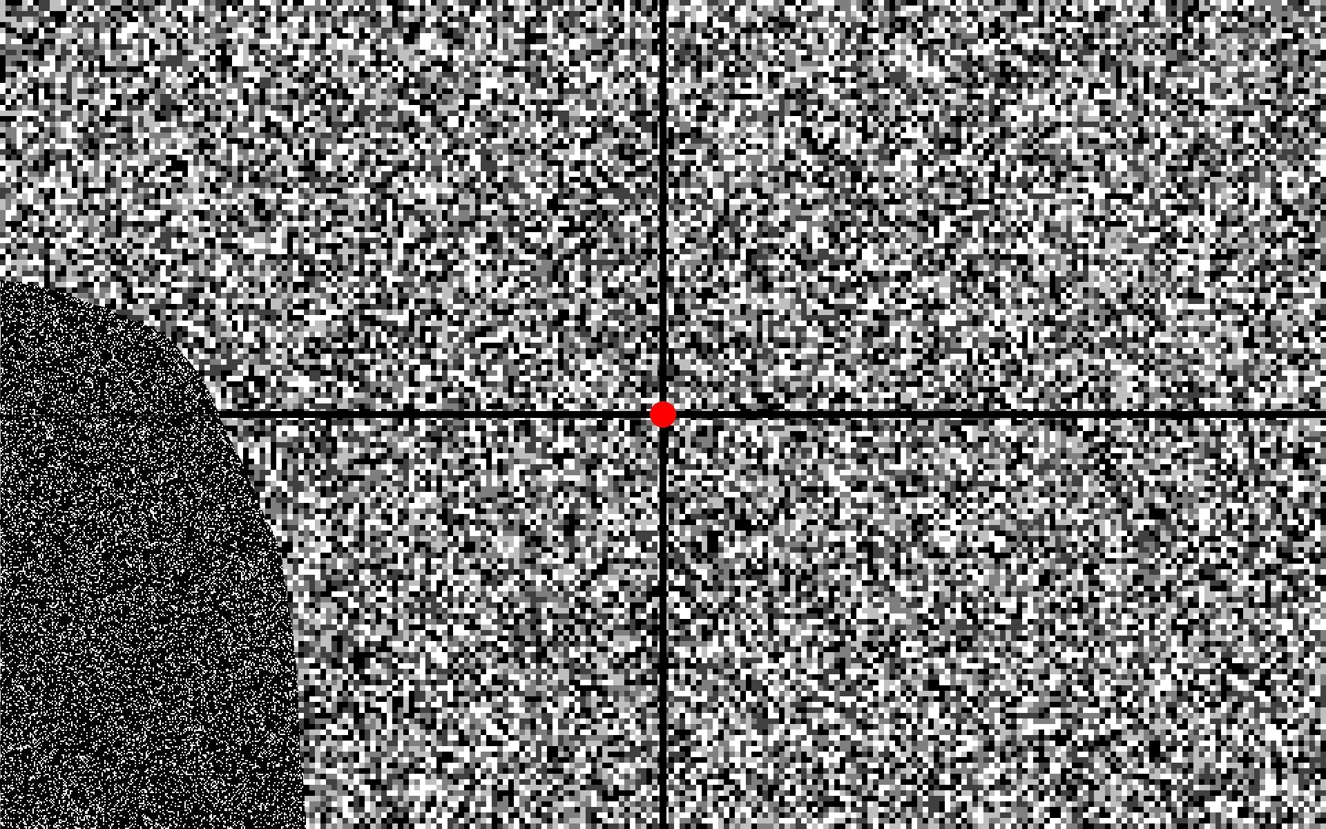
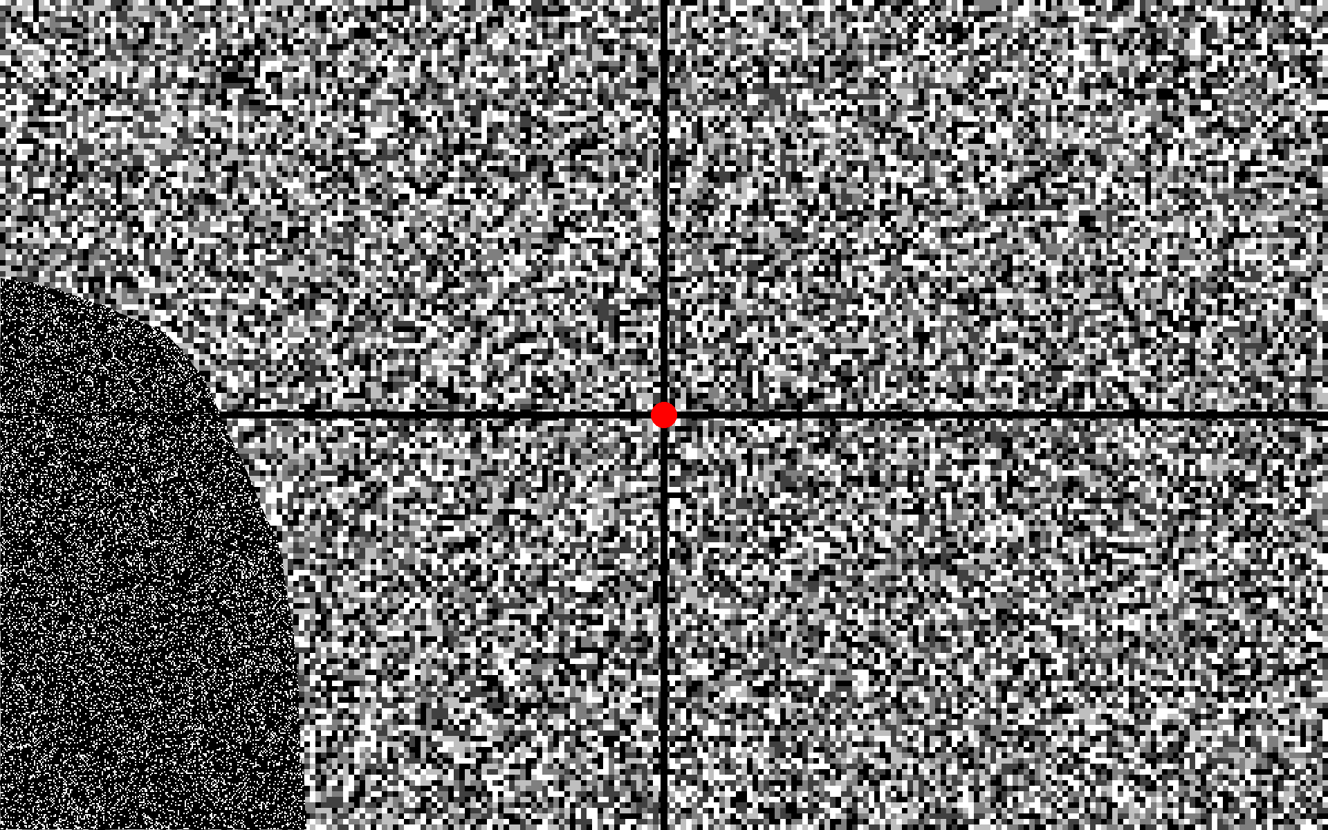
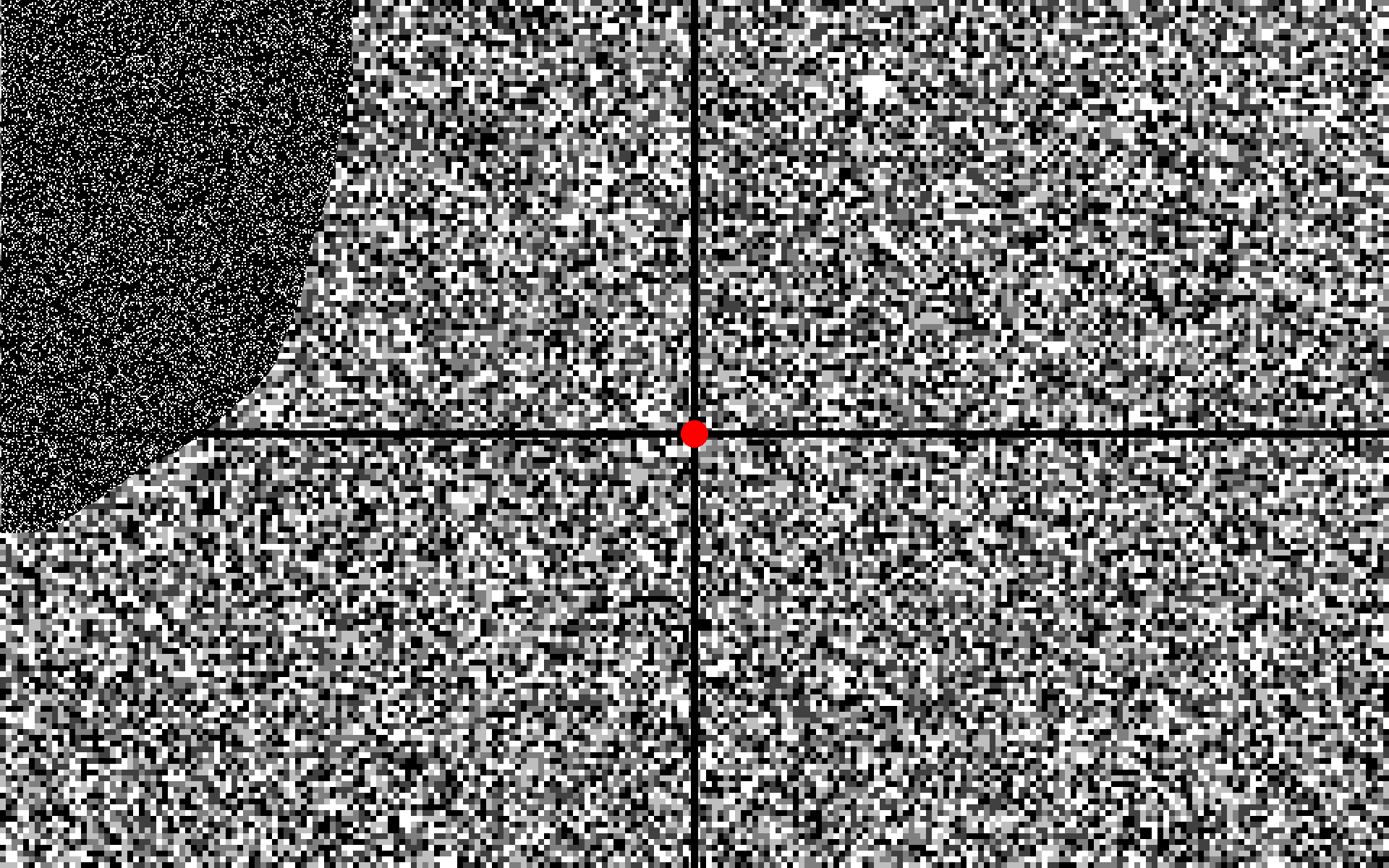
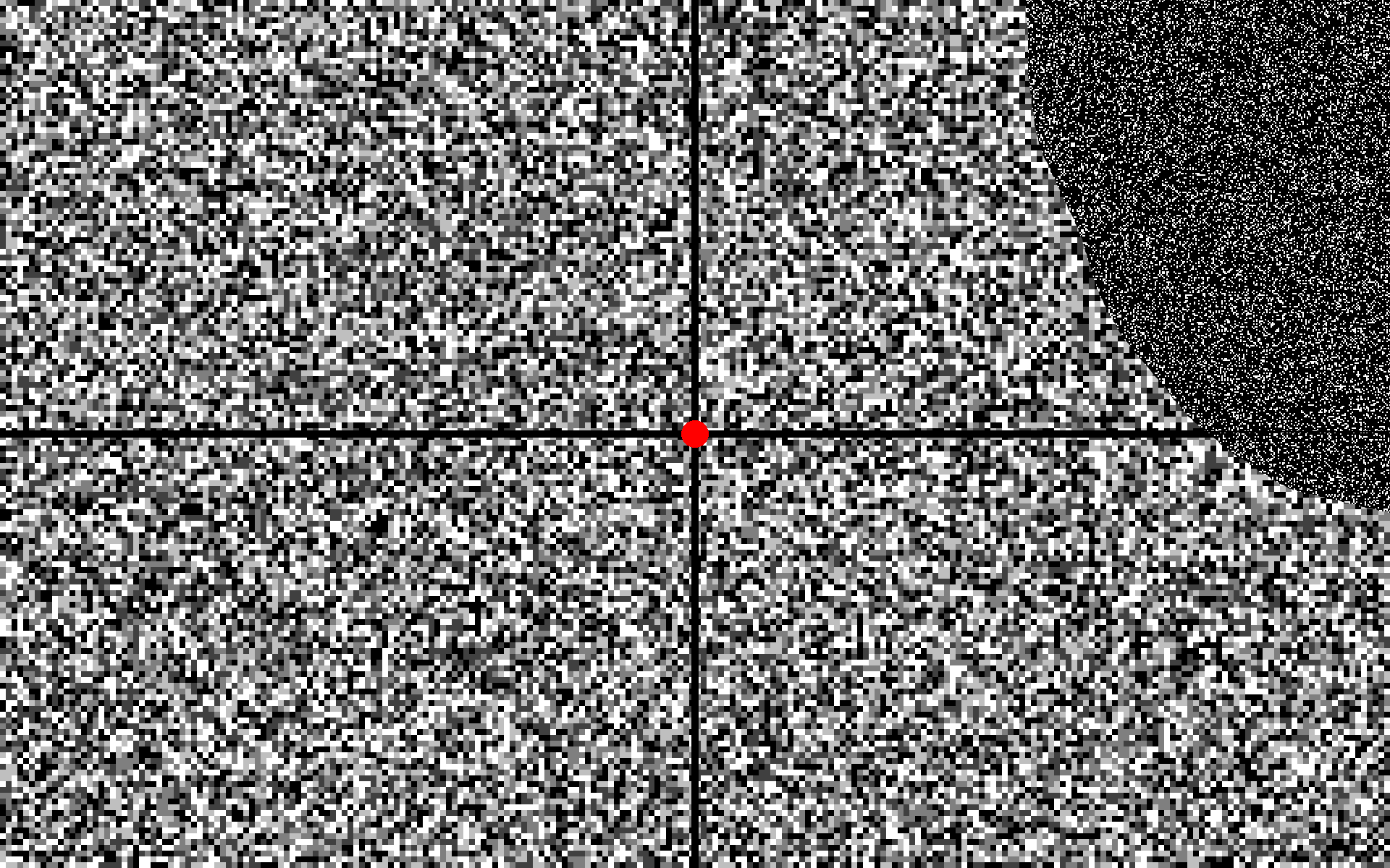
Horizontal and vertical separators divide the test image into four quadrants. Check for differences between the quadrants. Does the noise appear the same across the entire image, with no area looking different? That is a normal result. ⇒ Now test the other eye.
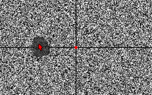
【Example 1】
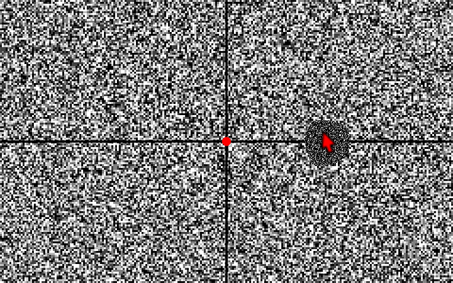
【Example 2】
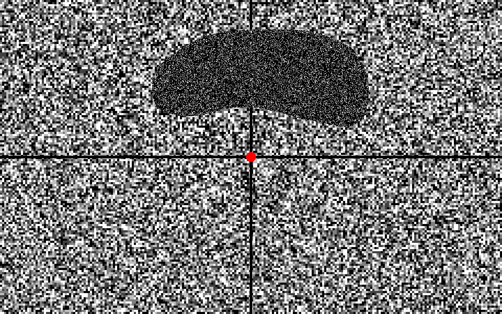
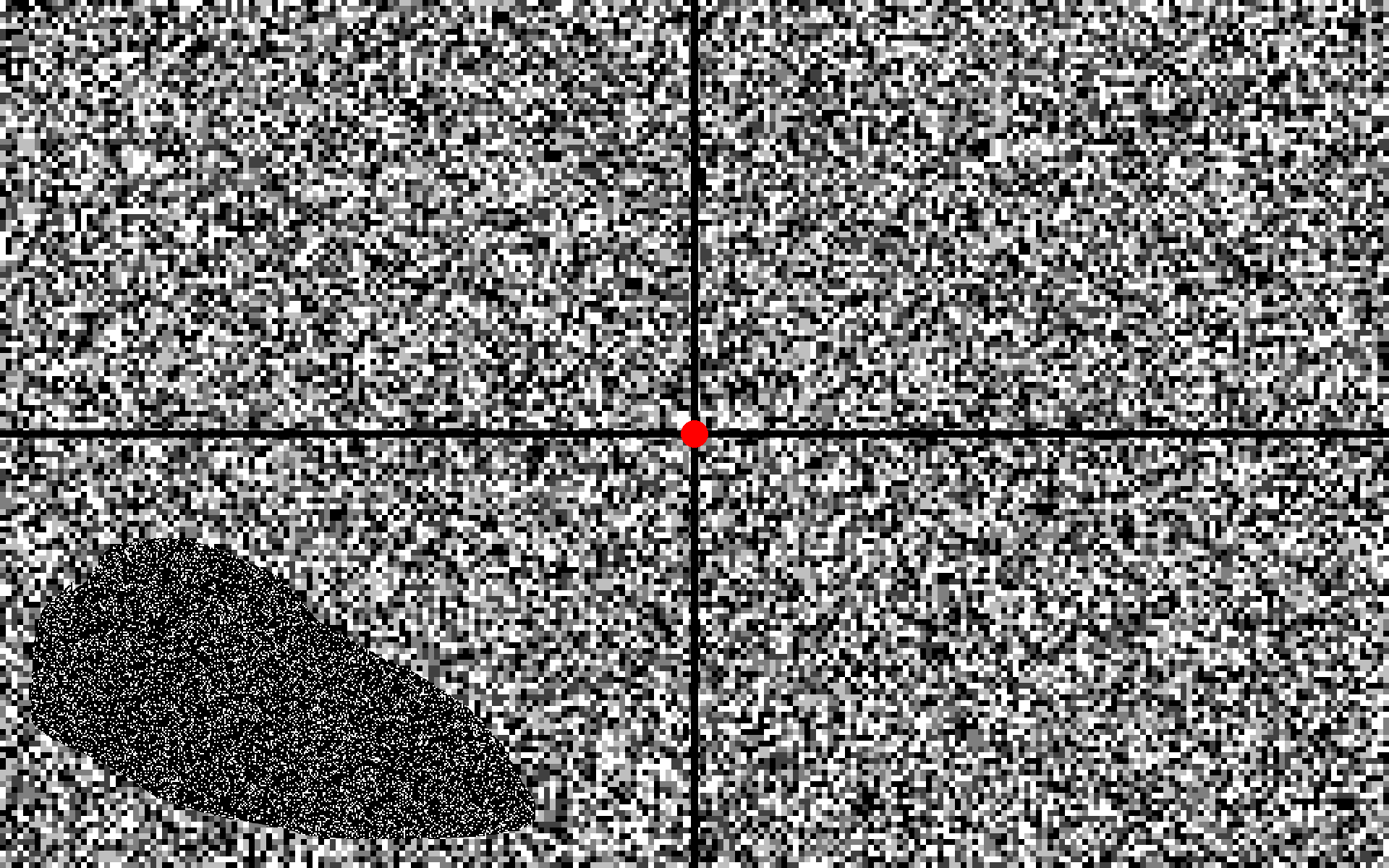
Do areas in the image appear different from other areas (“no flicker,” “little flicker,” “cloudy,” or “blackish/gray/grayish/white”)? Such areas are called “shadows” for convenience.
Alternate between the right and left eyes to determine if there is a difference in the shadows.
If shadows are seen, check their relationship with the fixation point and the quadrant separators.
Try changing the size of the noise particles and comparing the appearance of the shadows.
The size of the particles may affect the clarity of the shadows.
Compare what you see on the noise-moving image with what you see on a white and a black still image. Are the shadows more obvious on the noise-moving image than on the still images? Are the shadows always visible in the same location?
☆Any abnormality will be detected within a few seconds.
The test has no time limit, but 5 to 10 seconds is sufficient.
Understanding the results
If shadows always appear in the same location, a disease may be present.
Lack of shadows is normal; however, it is possible that the test may fail to detect abnormalities even if a disease is present.
×
The primary purpose of this web test is to help patients understand their medical condition.
Even in patients with a disease, the web test may not detect any abnormality.
Whether the results of the web test show abnormalities or not, please be sure to consult an ophthalmologist if you have any concerns.
Do you consent to begin the test?
No
Having consented, please select your display screen type:
※Not available for smartphones.
Special cases:
The red fixation point is not visible.
Serious eye disease is a possibility. We recommend that you schedule a consultation with an ophthalmologist as soon as possible.
Please note that this recommendation does not apply to patients who have already received ophthalmologic care and consented to the results.
A shadow is visible, but it moves and is not constant.
View the white still image. If the shadow appears more prominent on the white still image than on the noise-moving image, the shadow is highly likely to be a floater. Typically, when an individual with a visual field defect views the white still image, they report that either “nothing is felt” or “shadows are faint.”
Please note that we recommend a detailed ophthalmologic examination if a floater is suspected.
The noise flickers, and another noise becomes visible.
If you watch the noise-moving image for more than 10 seconds, a motion illusion may appear. Please alternate your view between the black still image and the noise-moving image or change the size of the noise particles. The illusion then usually disappears.
A circular shadow is noted toward the ear side on the horizontal separator.
You may be sensing Mariotte’s blind spot, which can occur in only one eye or in both eyes. The size of the shadow may differ between the left and right eye. This shadow could be pathological or within normal limits.
【Example 3】Mariotte’s blind spot
【Left eye】
【Right eye】
Move the red mouse pointer to the area where you see the shadow. Return your concentration to the fixation point. If you cannot see the red pointer in the shadow, then you are sensing Mariotte’s blind spot. If you move the red pointer into any other perceived shadow, you may not see the red pointer.
Note: Marriotte’s blind spot coincides with the optic nerve disc and is present even in a healthy person. It is usually unnoticeable.
Shadows are visible to both the right and the left eye on the same side, or the patients cannot see anything or feel that nothing is visible on the same side.
This defect on the same half (right or left) of each eye’s visual field is called homonymous hemianopia. An occipital lobe cerebral infarction (ischemic stroke) or another abnormality at the back of the head is a possibility. If the visual field defect is very large, the patient cannot recognize shadows and may feel that nothing can be seen in that direction.
In the case of an occipital lobe cerebral infarction, a sudden loss of vision in one direction occurs. Please visit a medical institution immediately.
【Example 4】Left inferior homonymous hemianopia from a right occipital lobe cerebral infarction.
【Left eye】
【Right eye】
A shadow appears toward the ear side in both the right and the left eye.
An abnormality in the optic chiasm (the area in the brain where the left and right optic nerves cross), such as a pituitary tumor, is a possibility.
【Example 5】Bitemporal superior quadrant hemianopia from a pituitary tumor.
【Left eye】
【Right eye】
In glaucoma, the nasal side of the image is often abnormal, and the right and left eyes often do not coincide.
We recommend that individuals observing shadows potentially associated with the foregoing diseases promptly seek medical attention.
Difference between the official and web versions of noise-field perimetry:
The official version of this test used in our clinic projects the noise-moving image directly to the display screen using computer graphics. The web version utilizes a video file converted from the noise-moving image, resulting in slightly lower quality and randomness than the official version.
References
Inoue A, Koike E, and Matsumoto C: Subjective perception of visual field defects using random noise-moving images—A comparison of CG noise and analog noise. Abstracts of the 9th Annual Meeting of the Japan Imaging and Perimetry Society, 58, 2020.
Inoue A: Noise-field campimetry. In: Negi A et al. (eds): Ophthalmologic Examination Guide. 3rd ed. Bunkodo, Tokyo: pp. 287–290, 2022 [Japanese].
Inoue A: Noise-Field Test. In: Matsumoto C. (eds): New Practical Ophthalmology 13. Visual Field Frontier. Bunkodo, Tokyo: pp. 83-88, 2024 [Japanese].
Inoue A, Koike E, Maeda N, Matsumoto C. Subjective perception of visual field defects using random noise-moving images in patients with glaucoma: A comparison of computer graphics and analog noises. PLoS One. 2024 May 20;19(5):e0303849. Open Access Journal PLOS ONE
×
The primary purpose of this web test is to help patients understand their medical condition.
Even in patients with a disease, the web test may not detect any abnormality.
Whether the results of the web test show abnormalities or not, please be sure to consult an ophthalmologist if you have any concerns.
Do you consent to begin the test?
No
Having consented, please select your display screen type:
※Not available for smartphones.